It’s important to monitor your health so you know if you are healthy. Knowing the status of your health can show you what to change in your diet, or what to heal.
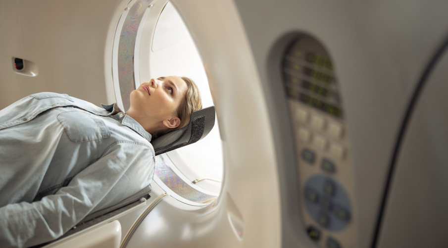
Body scanning diagnostic procedures, such as CT scan, ultrasound, MRI, and x rays are used to detect abnormalities in a body part or organ.
The rapid changes in medical technology have given birth to more advanced diagnostic tools, providing more detailed images and accurate data, such as the size of a tumor or possible factors causing a blockage in a blood vessel or numbing of a nerve.
But, what are the most common body scans used nowadays and how are they different from one another?
This article will tackle this topic for your increased awareness and understanding on available body scan options.
Because some people are afraid of the technology, and they might avoid having a check, this article will provide knowledge to help ease that fear or anxiety. Because when you know something you fear it less. And this can help people do check ups when they need to.
4 Common Body Scans:

1. Computerized Tomography Scan or so called CT Scan.
Through the use of CT scanner, medical practitioners are able to get clear images of the internal organs, blood vessels, and tissue structures of the human body.
It also produces images of the bones, arteries, neurological disorders, and heart abnormalities.
CT scan is used to identify problems, like blood clotting problems, abnormalities in the bone joints, head injuries, and brain tumor.
CT scanners work on the principle of using x ray in combination with software, which produces the images.
There are some images that are taken using x ray only, while some are created using software.
CT scanners have various options available for controlling the number of slices and the number of pictures to be taken.
The images produced by CT scanners are very clear, crisp, and high resolution. In addition to these, they are also portable.
Here’s how CT scan is different from other body scans:
Produces crispier and more detailed images.
CT scan has brought a revolutionary change in diagnosing the problems of patients, with the capability of producing detailed images of the internal organs.
It makes use of low level radiation to eliminate the effects of ionizing radiation, which are associated with x ray.
Low dose CT (computed tomography) can be used to prevent undue exposure to radiation, which can increase risk of cancer.
Diagnoses a wider range of medical conditions.
CT scans can be used to diagnose a wide range of health conditions, which can’t be diagnosed using magnetic resonance imaging and other methods.
Easier to use and more affordable.
CT scans have a lot of benefits over magnetic resonance imaging, such as reduced motion, and it is a less invasive procedure.
They allow the physicians and the nurses to move freely and focus on the important areas that need to be checked.
2. Magnetic Resonance Imaging or so called MRI.
MRI is a medical imaging procedure and technology that uses an extremely strong magnetic field, radio waves, and an elaborate computer to create detailed images of the internal organs, soft tissues, bones, and practically all other internal structures.
An MRI is basically a magnetic wave instrument that uses the right magnetic fields required to produce the detailed images needed by doctors.
MRI scanners are designed to capture a faint image of the target area by creating a very strong magnetic field around it.
The strength of the magnetic field depends on the mass of the object to be scanned. You may book a private MRI anytime.
Here’s how MRI scan is different from other body scans:
No harmful ionizing radiation.
Unlike x rays or CT scans, an MRI does not utilize harmful ionizing imaging radiation.
Uses radio waves.
While CT scan and x rays use electromagnetic radiation, MRI uses strong magnetic fields and radio waves to create images of the internal organs or structures of the human body.
Not applicable for those with pacemakers.
MRI has a powerful magnetic field that can cause damage to internal metal devices, like a heart pacemaker. Unlike MRI, patients with pacemakers or cardioverter defibrillators may undergo CT scan without causing problems.
3. Ultrasound.
Ultrasound is an electrical form of energy that uses waves of high frequency.
It’s often used to produce an image of the internal body structures, like tendons, bones, muscles, and arteries.
The main goal of ultrasound is to locate or eliminate pathology without causing any damage to internal structures.
The main uses of ultrasound are for pregnancy sonograms, vascular, cellular, facial cosmetic and breast enhancements, gynecological inspections, anesthesia uses, radiology, and bone growth analysis, among many others.
Diagnostic ultrasound uses sound waves to produce images of internal abnormalities, which are compared to what’s observed in real life with an x-ray.
Here’s how ultrasound is different from other imaging methods:
Use of soundwaves.
These high frequency sound waves do not require any extra equipment or contraptions.
They usually pass straight through the body without hitting any obstacles or internal structures.
These sound waves are not able to be heard by humans as they normally come in at frequencies below 1000 hertz.
The human ear has a limited ability to detect these low frequencies.
Applications.
While CT and MRI scans are widely used to detect tumor and other severe medical conditions, like cancer, ultrasound is commonly used for detecting pregnancy, as well as kidney, uterine, and bladder problems.
4. Digital X Rays.
Digital radiography or x ray is a new type of radiography that utilizes x ray receptors, directly capturing data during the medical patient’s examination.
This technology is rapidly replacing the older forms of radiographic imaging, such as MRI and ultrasound.
Digital dental x rays have helped dentists save money by letting them detect problems earlier and developing preventative treatments.
Is digital x ray radiation safe?
Yes, because it causes very little damage to tissues and does not penetrate the bones.
In short, digital x rays have been established as safe for dental use.
Here’s how digital x-ray is different from other body imaging scans:
Faster.
Easier to do.
More affordable.
More applicable for dental, bone, and lung problems.
If You Put All In Comparison:
Body imaging methods, such as CT scan, MRI, ultrasound, and x rays are used in detecting various medical conditions.
X rays are usually used in detecting dental, bone, and lung problems, such as fractures and bronchitis.
Cancer or tumor growths can usually require more detailed and crispier images, like what MRI and CT scans provide.
On the other hand, ultrasound is usually used in diagnosing pregnancy, gynecologic problems, as well as kidney and uterine problems.
